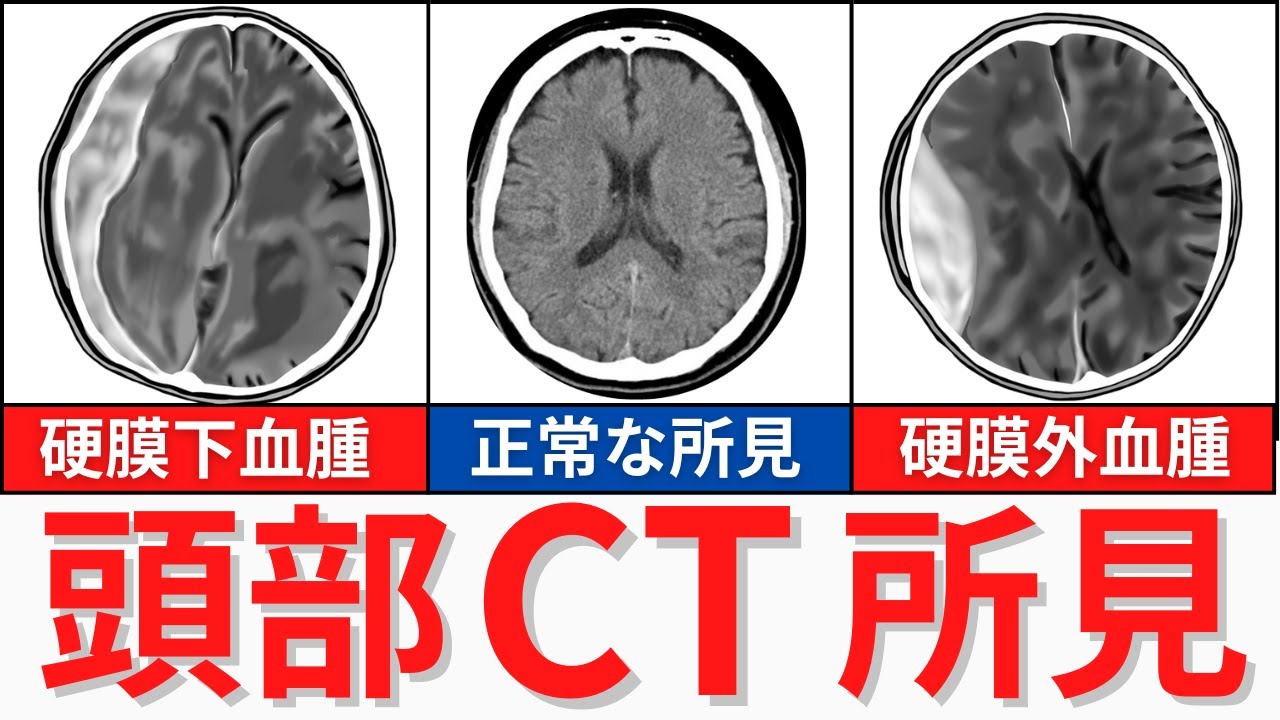
【10分で解説】CT検査の所見について解説
Keywords
Summary
183 words
Critical Evaluation
Value of the Information & Strength of the Argument
The video provides valuable information for nursing students, clearly explaining the radiological features of three important intracranial hemorrhages. The argumentation is solid, as the presenter logically connects anatomy, pathophysiology, and imaging findings. He uses analogies (e.g., ‘convex lens’ vs ‘crescent’) to aid memorization and reinforces learning with practice questions. The explanations are consistent with standard medical knowledge, and the presenter emphasizes the clinical significance of each finding. However, the video does not delve into differential diagnoses or advanced imaging techniques, but for its target audience, it is highly effective.
Scientific Rigor, Source Quality, Title Accuracy
The scientific rigor is adequate for an educational video aimed at nursing students. The presenter does not cite specific sources, but the information aligns with established medical textbooks and guidelines. The title accurately reflects the content, though the ‘10 minutes’ is misleading as the video is over 33 minutes long. The video includes practice questions from national nursing exams, which adds credibility. The description provides links to related videos and social media, but no direct references to scientific literature. Overall, the content is reliable for its intended purpose.
192 words
Title / Content Match
The title accurately reflects the content, which focuses on interpreting CT findings, though the '10 minutes' is misleading as the video is over 33 minutes long.
Quality & Reliability
8/10
The video provides a clear and structured explanation of head CT findings for subarachnoid hemorrhage, subdural hematoma, and epidural hematoma, integrating anatomy and symptoms. The content aligns with standard medical knowledge, and the presenter uses a pedagogical approach. However, the video is primarily educational for nursing students and does not cite primary sources directly, relying on established medical facts.
Chapters
Cited Sources
- Related video: Subarachnoid hemorrhage explanation — Mentioned in the description as a related video for further explanation of subarachnoid hemorrhage.
- Related video: X-ray findings explanation — Mentioned in the description as a related video for X-ray findings.
- Recommended playlist: Important anatomy, diseases, and treatments — Mentioned in the description as a recommended playlist for further study.
Concurring Sources
- Subarachnoid hemorrhage - Wikipedia — Confirms the CT findings and symptoms described in the video.
- Epidural hematoma - Wikipedia — Confirms the biconvex appearance on CT and the mechanism of bleeding.
- Subdural hematoma - Wikipedia — Confirms the crescent-shaped appearance on CT and the pathophysiology.
Contribution & Novelties
The video offers a clear and systematic approach to interpreting head CT findings for three common hemorrhagic conditions, which is particularly useful for nursing students. It integrates anatomy, pathophysiology, and clinical presentation, making it a comprehensive educational resource. The use of mnemonics and practice questions enhances retention.
Pour aller plus loin :
- Subarachnoid hemorrhage - Wikipedia — Provides a detailed overview of the condition, including causes, symptoms, and diagnosis.
- Epidural hematoma - Wikipedia — Explains the pathophysiology and imaging characteristics of epidural hematomas.
- Subdural hematoma - Wikipedia — Discusses the types, symptoms, and radiological findings of subdural hematomas.
- Cushing reflex - Wikipedia — Describes the physiological response to increased intracranial pressure, which is mentioned in the video.
117 words
Radar Profile
The radar profile shows high scores in information quantity, quality, and reliability, with a slightly lower technical level, indicating that the video is well-suited for its target audience of nursing students, providing comprehensive and accurate information without excessive technical complexity.