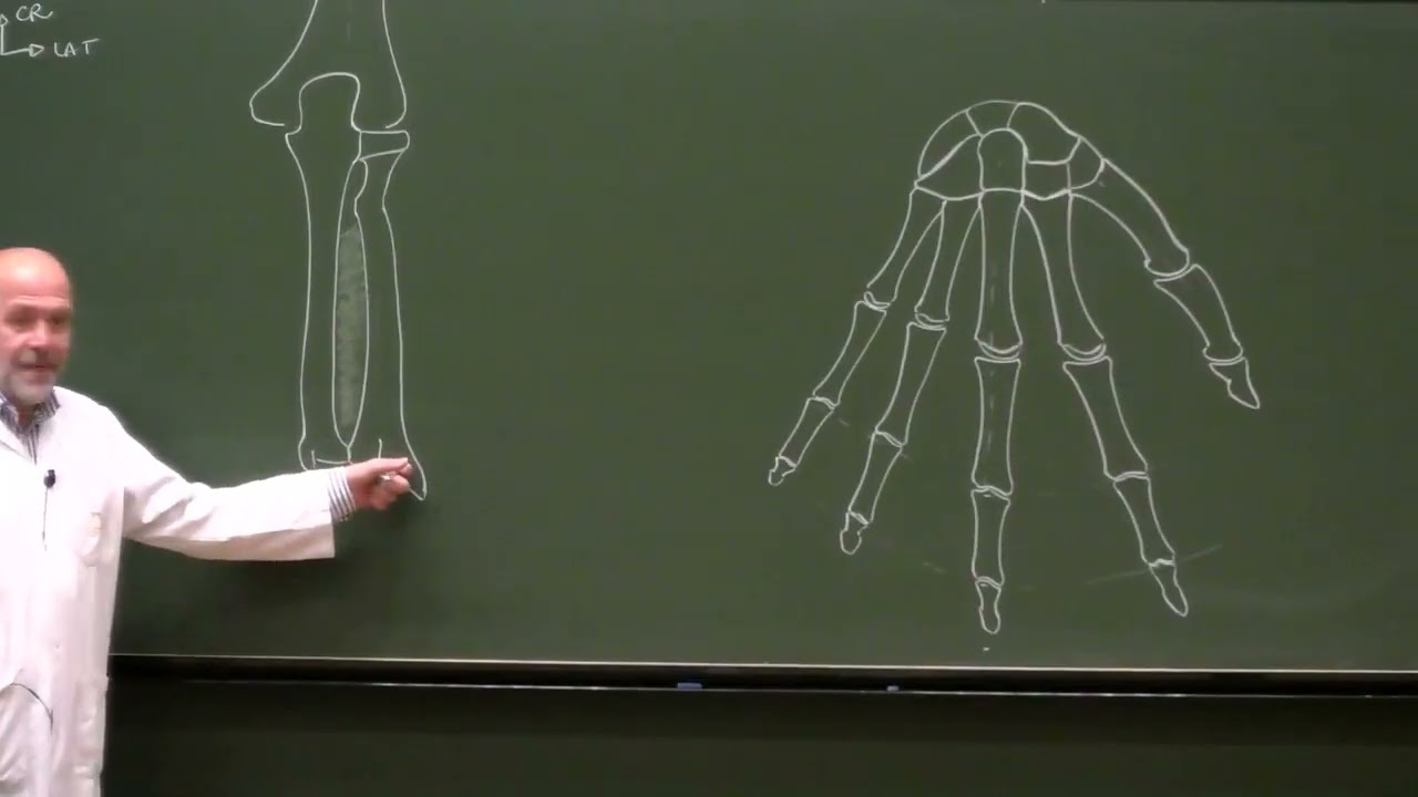
Muscles de l'avant-bras, 2ème partie : compartiment postérieur (version 2026)
Muscles of the forearm, 2nd part: posterior compartment (2026 version)
Title history
- Muscles de l'avant-bras, 2ème partie : compartiment postérieur (version 2026) — observed on 20261112-1339 UTC (YouTube)
Keywords
Summary
177 words
Critical Evaluation
Value of the Information & Strength of the Argument
The educational value is exceptionally high for a specialized audience, presenting complex anatomical data in a clear, step-by-step drawing methodology. The argumentation relies on established anatomical consensus, with rigorous explanations that connect spatial relationships (e.g., tendon spacing, compartmentalization) to functional consequences, such as the mechanical advantage provided by the extensor retinaculum. The instructor effectively uses repetition and mnemonic anchors (e.g., ‘head of radius proximal, head of ulna distal’) to solidify understanding without sacrificing scientific precision.
Scientific Rigor, Source Quality, Title Accuracy
The scientific rigor is solid, reflecting standard instructional protocols in medical education. The content aligns perfectly with contemporary anatomical terminology, and the reference to the historical anatomist Léo Testut is contextualized, noting the evolution from his classification of the anconeus to current independent muscle status. The title ‘Muscles de l’avant-bras, 2ème partie : compartiment postérieur’ is a faithful indicator of the video’s scope, leaving no ambiguity about the subject matter. No comments were provided for analysis.
166 words
Title / Content Match
The title perfectly matches the content, which exclusively covers the posterior compartment of the forearm as the second part of a dedicated series.
Quality & Reliability
9/10
The lecture is methodical, based on current anatomical nomenclature, and clearly distinguishes the superficial and deep layers with precise origins, insertions, innervations, and actions. The reference to Leo Testut provides historical context without compromising modern classification accuracy.
Key Moments
Markers derived by PSI from the transcript: the creator did not define chapters.
- Presentation of the dorsal osseous view (radius, ulna, carpal bones) and the interosseous membrane.
- Introduction to the superficial layer: common extensor origin and detailed description of the extensor digitorum muscle.
- Analysis of the extensor digiti minimi, its independent osteofibrous canal, and its fusion with the common extensor tendon.
- Study of the extensor carpi ulnaris (two heads) and the anconeus muscle, referencing its historical classification by Testut.
- Deep layer begins: origins, courses of the abductor pollicis longus and extensor pollicis brevis, sharing a common compartment.
- Extensor pollicis longus, its reflection on the dorsal tubercle, and the delineation of the anatomical snuffbox.
Cited Sources
- Léo Testut's anatomical works — Mentioned as a historical reference for the classification of the anconeus muscle, which he proposed to include in the triceps brachii (quadriceps brachii).
Concurring Sources
- Gray's Anatomy for Students — Standard anatomical textbook describing the same posterior compartment muscles, origins, and innervation with consistent nomenclature.
Dissenting Sources
- Testut's historical classification — Leo Testut historically grouped the anconeus muscle with the triceps brachii, whereas the current anatomical nomenclature recognizes it as an independent muscle of the posterior forearm compartment.
Contribution & Novelties
The apport of this video is its highly structured didactic methodology, offering multiple dorsal views and a progressive construction of muscle layers that clarifies complex spatial arrangements—crucial for students and clinicians. While the factual content is standard anatomy, the pedagogical clarity and explicit linking of structures to clinical signs (like the snuffbox) constitute a valuable educational contribution. The historical note on Testut provides insight into the evolution of anatomical classification.
Pour aller plus loin :
- Extensor digitorum muscle — Core muscle of the superficial posterior compartment, detailed origin and insertions directly relevant to the lecture.
- Extensor carpi ulnaris muscle — Key superficial muscle with two heads, central to wrist extension and ulnar deviation.
- Anatomical snuffbox — The clinical landmark mentioned at the end of the video, formed by the thumb tendons.
- Radial nerve — The primary innervation of all muscles in this compartment, crucial for understanding functional deficits.
148 words
Radar Profile
Le profil radar montre un polygone extrêmement équilibré avec des scores élevés (9/10) sur les quatre axes : quantité d'information, qualité, niveau technique et fiabilité. Ce profil traduit une ressource pédagogique dense, robuste et hautement spécialisée, adaptée à un public étudiant avancé.